Private Label, White Label, Wholesale partnerships available - EU, USA and UK - Free shipping from €75
Mechanism of Peptide Bond: How Amide Links Form and Break
A peptide bond is an amide link formed when the carboxyl group of one amino acid condenses with the amino group of the next, releasing one molecule of water. Yet cells build this bond by transferring an activated acyl group, not by squeezing water out of free amino acids.
That distinction explains an apparent contradiction: a peptide backbone can persist in a sealed aqueous vial while the ribosome rapidly extends a growing protein. The difference lies in the starting materials, leaving groups, and reaction barriers, not in a special kind of peptide bond.
By Herbilabs
Table of Contents
- The Short Answer on Peptide Bond Chemistry
- Peptide Bond Formation and Cleavage Compared
- Why the Peptide Bond Is Planar and Partly Double
- How Free Amino Acids Link Without an Enzyme
- Indirect Transesterification and O-to-N Routes
- Ribosomal Aminolysis Step by Step
- Where the Ribosome's RNA Catalyst Lowers the Barrier
- How Peptide Bonds Break and Why They Last
- What to Control When Working With Peptides in the Lab
The Short Answer on Peptide Bond Chemistry
Consider glycine joining alanine. The resulting backbone contains a C(=O)–N amide linkage, with the carbonyl carbon from one amino acid joined to the nitrogen of the next.
A simple analogy is two halves of a zipper locking together. The joined teeth represent the new connection, while water represents the small piece formally removed during condensation. The analogy describes the overall accounting, however, not every chemical step.
Formation connects amino acids into chains; hydrolysis incorporates water and cleaves the linkage. The useful distinction is among these routes:
- Flask condensation: Free amino acids generally need chemical activation before productive coupling.
- Ribosomal acyl transfer: An incoming amino group attacks an already activated substrate.
- Protease cleavage: An enzyme makes selected backbone bonds accessible to hydrolysis.
The peptide bond examples from Herbilabs help connect the structural formula to recognizable molecules. For readers approaching peptides through skincare, Skin Perfection's resource on plant-derived oils for facial care provides context for a different ingredient class; oils and peptides shouldn't be assumed to share the same chemistry.
Understanding the mechanism of peptide bond formation requires both a reaction comparison and a closer look at intermediates. For laboratory handling, activation barriers and storage controls matter just as much as the final molecular structure.
Peptide Bond Formation and Cleavage Compared
An electrophile is the electron-poor reaction center, usually the carbonyl carbon in these reactions. A nucleophile supplies the electron pair that forms a bond to that center.
Peptide bond formation and cleavage routes compared
| Route | Electrophile | Nucleophile | Driving force | Practical speed |
|---|---|---|---|---|
| Non-enzymatic condensation | Activated carboxyl, such as an ester or active intermediate | Free alpha-amino group | Coupling reagent or chemical activation | Slow without activation |
| Uncatalysed hydrolysis (cleavage) | Amide carbonyl | Water | Thermodynamically favourable | Very slow under ordinary conditions |
| Ribosomal peptidyl transfer | Peptidyl-tRNA ester carbonyl | A-site aminoacyl-tRNA alpha-amino group | Pre-activated ester positioned by rRNA | Fast on the intact ribosome |
| Protease cleavage | Amide carbonyl | Water activated by catalytic residues | Enzyme stabilises the transition state | Fast and sequence-specific |
For non-enzymatic condensation, activation creates a carbonyl partner that an amine can attack productively. Mixing amino acids in water usually produces an acid-base mixture, not a useful peptide synthesis.
Uncatalysed hydrolysis illustrates the difference between a favourable outcome and an accessible pathway. Products can be energetically favourable even when the reaction takes place too slowly to matter during routine handling.
Ribosomal peptidyl transfer starts with energy already invested in an activated substrate. Proteases instead accelerate cleavage by providing a catalytic environment, although some proteases use an enzyme nucleophile before water completes the reaction.
Reading rule: Activation and catalysis change the pathway and speed. A catalyst doesn't change the reaction's equilibrium.
The table deliberately leaves out numerical rate constants and detailed transition-state structures. Neither a universal synthesis speed nor a universal peptide lifetime can be assigned from the bond formula alone.
For any proposed peptide reaction, the useful questions are concrete: What is the electrophile? What is the leaving group? What controls the speed?
Why the Peptide Bond Is Planar and Partly Double
An ordinary single bond resembles a freely turning hinge. The peptide C–N bond is more like a hinge reinforced by a shared brace: the nitrogen's lone-pair electrons overlap with the neighbouring carbonyl system.
The molecular model highlights the resulting flat peptide unit.
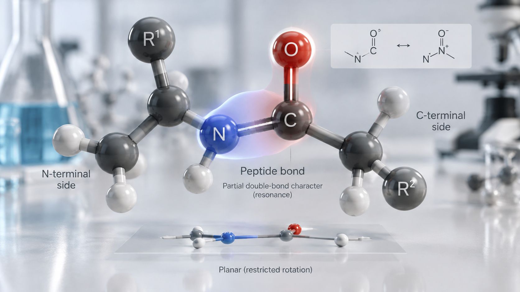
This electron sharing is resonance, not a rapid alternation between separate molecules. The nitrogen lone pair delocalizes into the carbonyl π* system, so a drawing containing only a C–N single bond doesn't fully describe the electronic structure.
What resonance changes
The C–N bond becomes shorter and stronger than a typical amine C–N bond, and rotation becomes restricted. The NCBI Bookshelf peptide-bond overview describes partial double-bond character and the approximately planar arrangement of the six atoms comprising the peptide unit: the adjacent alpha carbons, carbonyl carbon and oxygen, nitrogen, and its attached hydrogen.
That last detail can be confusing in condensed drawings. The oxygen belongs to the planar unit even when a backbone sketch places it above the main chain.
- Restricted rotation: Twisting the amide disrupts the favourable overlap of the nitrogen and carbonyl orbitals.
- Trans preference: For most peptide bonds, placing neighbouring backbone groups on opposite sides reduces steric crowding compared with the cis arrangement.
- Remaining flexibility: Rotation occurs mainly around the N–Cα and Cα–C(O) bonds, described by the φ and ψ torsion angles.
The animation offers another way to follow the backbone geometry.
A chain of plates and hinges
A polypeptide is better pictured as linked rigid plates than as a uniformly floppy string. The peptide units are the plates; the neighbouring backbone bonds supply the hinges.
Those hinges can't adopt every imaginable angle because atoms occupy space. Hydrogen bonding, side-chain chemistry, and the surrounding solvent further narrow the accessible conformations, so the sequence doesn't specify a unique shape independently of conditions.
Urea provides a practical distinction between unfolding and cleavage. It can disrupt the interactions supporting a protein's higher-order structure without readily cutting the covalent backbone. An unfolded protein therefore isn't automatically a hydrolysed protein, and an intact backbone doesn't guarantee preserved biological activity.
How Free Amino Acids Link Without an Enzyme
A flask containing amino acids and water lacks a convenient way to replace the carboxyl group's hydroxyl with an amino nitrogen. Under many useful conditions, the amino group is protonated and the carboxyl group is present as a carboxylate, neither an ideal pairing for direct amide formation.
Even when the free acid is present, its carbonyl isn't sufficiently reactive for efficient coupling, and hydroxide is a poor leaving group. Direct dehydration may require harsh conditions that are unsuitable for sensitive amino acids.
Activation supplies a workable leaving group
Activation resembles attaching a sound handle to a suitcase with a broken one. The contents haven't changed, but a previously awkward operation becomes manageable.
Chemists can convert a carboxyl group into an activated ester or, where appropriate, an acyl chloride. The amine attacks the activated carbonyl, produces a tetrahedral intermediate, and displaces the leaving group as the carbonyl reforms.
Carbodiimides provide another common route:
- DCC activation: Dicyclohexylcarbodiimide converts the carboxyl group into an O-acylisourea intermediate that can undergo amine attack.
- EDC coupling: A related carbodiimide provides an activation option often chosen for workflows requiring different solubility and work-up properties.
- Product formation: Productive substitution gives the amide and a reagent-derived urea by-product, rather than ejecting water from unactivated amino acids.
Activation doesn't guarantee clean conversion. Competing hydrolysis and unproductive rearrangement of activated intermediates can consume material before the desired amine reacts.
Keeping chain growth selective
In solid-phase peptide synthesis, a growing peptide remains attached to a resin while activated amino-acid building blocks are added sequentially. Fmoc or Boc protecting groups prevent unwanted reactions at amino groups, and suitable side-chain protection limits branching or other coupling errors.
Two problems deserve particular attention. Activation can promote racemisation at the alpha carbon, so coupling conditions must preserve stereochemistry rather than merely produce the expected mass.
Water also competes with the intended amine for susceptible activated intermediates. For direct condensation, aqueous conditions generally favour hydrolysis; activated coupling in water can still work when reagent choice and reaction conditions support it.
A clean mass signal doesn't establish correct stereochemistry. Coupling quality includes both connectivity and configuration.
Indirect Transesterification and O-to-N Routes
Direct aminolysis isn't the only possible route to an amide. An indirect pathway first places the acyl group on oxygen, then transfers it to nitrogen.
A 2025 Journal of the American Chemical Society study investigated transesterification followed by an O-to-N rearrangement using fluorine-tagged aminoacyl adenylate esters and 19F NMR kinetic monitoring. The fluorine label supplied a spectroscopic handle for following the chemistry rather than serving as the new peptide linkage.
Following the acyl group
- Activated starting material: Aminoacyl adenylate esters provide reactive acyl donors.
- Oxygen-linked intermediate: Transesterification transfers the acyl group to an oxygen-containing acceptor, producing an ester intermediate.
- Nitrogen-linked product: An O-to-N acyl shift converts that oxygen-linked arrangement into the amide.
- Kinetic observation: NMR monitoring follows the reaction sequence so individual transformations can be distinguished rather than inferred solely from the final product.
The important contrast is the identity of the initial attacking atom. Direct aminolysis starts with nitrogen attack; the indirect route passes through an oxygen-linked species before amide formation.
Kinetic measurements can help identify which transformation limits overall conversion, but a particular rate-limiting assignment requires the original experimental results and conditions. This remains a research pathway under investigation, not a replacement for the established ribosomal mechanism or proof that the same route operates in cells.
Ribosomal Aminolysis Step by Step
During protein synthesis, the ribosome receives amino acids attached to transfer RNA, or tRNA. The growing chain is also tethered to tRNA, which makes the substrate chemically different from a free peptide carboxylic acid.
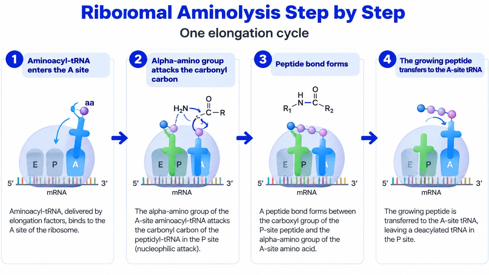
The PNAS structural account of peptidyl transfer describes attack by the A-site amino group on the ester carbonyl of the P-site peptidyl-tRNA, followed by transfer through a tetrahedral oxyanion intermediate. The large-subunit peptidyl-transferase center aligns the reacting tRNA acceptor ends.
The diagram summarizes the main chemical events within an elongation cycle.
From substrate delivery to chain transfer
An aminoacyl-tRNA enters the A site. It carries the incoming amino acid, while the P-site tRNA carries the growing peptide. Delivery matters because the ribosome doesn't collect unactivated amino acids and directly dehydrate them.
The alpha-amino nitrogen attacks the ester carbonyl. Its electron pair forms a new bond to the carbonyl carbon connecting the existing peptide to the P-site tRNA. The target is that activated ester carbonyl, not an internal amide somewhere along the growing chain.
A tetrahedral oxyanion intermediate forms. The former carbonyl carbon adopts a tetrahedral arrangement, and negative charge develops on oxygen. This is a chemical intermediate requiring stabilization, not merely a drawing of two amino acids touching.
The intermediate collapses and the peptide transfers. The carbonyl reforms while the linkage to the P-site tRNA is broken, leaving the elongated peptide attached to the A-site tRNA. The incoming amino acid becomes part of the chain without the ribosome generating water through direct condensation.
The former peptide carrier becomes uncharged tRNA. Peptide transfer removes its acyl attachment, but that doesn't mean the entire tRNA instantly leaves the ribosome. Subsequent movements carry the deacylated tRNA toward its exit pathway.
Translocation resets the arrangement. The peptidyl-tRNA moves toward the P site, and the A site becomes available for another incoming substrate. Translocation prepares the next reaction; it isn't the bond-forming chemical step itself.
Keeping chemistry separate from decoding
Messenger RNA determines which aminoacyl-tRNA is selected through decoding. The peptide-forming chemistry occurs at the acceptor ends of the tRNAs in the peptidyl-transferase center, not at the codon-anticodon contact itself.
The distinction also matters when laboratory terminology overlaps with clinical discussion. Cost Plus TRT's peptide injection guide addresses administration context; it shouldn't be treated as a ribosomal synthesis protocol or as authorization to administer research-use materials.
Each chemical cycle transfers an acyl group from an activated ester to an amino nitrogen. Cellular energy investment precedes this transfer through substrate preparation and supports the wider translation cycle; the reaction isn't powered by making free amino acids lose water on demand.
Where the Ribosome's RNA Catalyst Lowers the Barrier
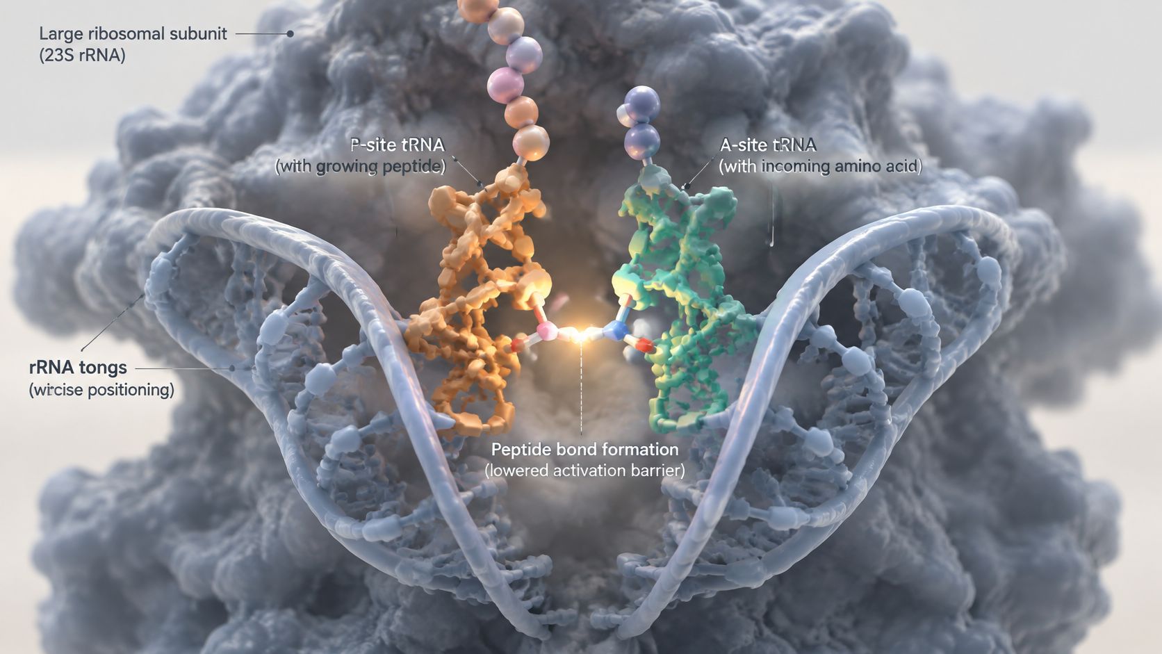
A pair of tongs can hold two pieces in a useful orientation, but the ribosome does more than hold substrates together. Its catalytic environment also organizes charge, hydrogen bonds, and proton movement during the reaction.
The molecular view locates that environment within the large ribosomal subunit.
An RNA-based catalytic toolkit
The ribosomal catalysis review hosted by PubMed Central describes substrate positioning, conformational organization, proposed proton shuttling involving the 2′-hydroxyl of terminal adenosine A76, and a coordinated water molecule that helps stabilize the oxyanion. These contributions belong to a reaction environment made primarily from RNA and the tRNA substrates, rather than a conventional protein-enzyme active site.
Correct alignment reduces the need for substrates to find a productive orientation by chance. Organizing the surroundings also helps accommodate the changing charge distribution as the reaction proceeds.
- Substrate positioning: The reacting ends meet in a geometry suitable for attack.
- Proton-transfer assistance: A proposed relay allows nitrogen and leaving-group protonation to change during bond rearrangement.
- Electrostatic stabilization: Local interactions support the negatively charged oxygen-containing intermediate.
The proposed proton shuttle shouldn't be read as a settled claim that a single group performs every catalytic task. Several coordinated interactions contribute to the overall barrier.
What the transition-state debate means
A kinetic-isotope-effect study of ribosomal peptide transfer contrasts a 2002 structural model describing a concerted process with a later interpretation in which intermediate formation and proton transfer occur together in the rate-limiting step, followed by faster, separate collapse.
In the concerted description, the major bond and proton rearrangements belong to a shared kinetic event. In the stepwise interpretation, the intermediate has a distinct place between the slower formation event and faster breakdown.
Both descriptions retain nitrogen attack, tetrahedral intermediate formation, proton transfer, and ester cleavage. Their disagreement concerns the energy profile and timing, not whether the ribosome presses free amino acids together.
A 2025 Journal of Biological Chemistry study reported that peptide bond formation and peptidyl release catalyzed by the isolated 50S subunit were more than 1,000-fold slower than the corresponding reactions catalyzed by the complete 70S ribosome. Under those experimental conditions, the complete particle was required for the faster rates; identifying the catalytic center doesn't make the rest of the ribosome dispensable.
How Peptide Bonds Break and Why They Last
Hydrolysis adds water across an amide linkage. The C–N bond breaks while new C–O and N–H bonds form, giving carboxyl and amino products whose protonation states reflect the solution conditions.
The obstacle is the reaction pathway. The carbonyl geometry, nitrogen bonding, proton positions, and surrounding water network must reorganize before cleavage can occur.
The peptide hydrolysis analysis hosted by PubMed Central reports uncatalysed activation energies of approximately 96–105 kJ/mol, consistent with the persistence of intact proteins during many ordinary aqueous handling conditions. This activation barrier describes access to the transition state, not the amount of energy released by the overall reaction.
Routes to faster cleavage
| Route or condition | What changes | Practical consequence |
|---|---|---|
| Acid catalysis | Carbonyl activation and proton-transfer pathways | Cleavage accelerates under suitable acidic conditions |
| Base catalysis | Availability of a stronger attacking nucleophile | Alkaline conditions can promote hydrolysis |
| Protease catalysis | Substrate alignment and transition-state stabilization | Selected accessible bonds can be cleaved efficiently |
| Local unfolding | Exposure of previously shielded backbone regions | Chemical or enzymatic access can increase |
Acid catalysis can improve electrophile activation and assist leaving-group protonation. Base catalysis supplies a more reactive nucleophile than neutral water, although the outcome still reflects the substrate and reaction conditions.
Neither route supplies a universal mild procedure for complete peptide cleavage. Conditions strong enough to force hydrolysis can also damage sensitive side chains or compromise the analytical purpose of the sample.
Enzyme access and local structure
Proteases use catalytic acid-base residues, metal ions, or covalent enzyme intermediates to lower the cleavage barrier. Their binding regions also recognize particular substrate features, so enzyme-mediated degradation isn't equivalent to random attack by bulk water.
A potentially cleavable sequence may remain protected inside a folded protein. Local opening or unfolding can expose it, allowing access before the bond-breaking chemistry begins.
Storage insight: A sealed vial limits external contamination, but it doesn't eliminate water, internal contaminants, or every degradation pathway.
Researchers who compare powder and solution stability should distinguish backbone cleavage from loss of usable material through aggregation, oxidation, adsorption, or other changes. A peptide can lose experimental suitability without extensive hydrolysis.
Practical stability therefore requires control of pH, temperature, time, and contamination. Treating every peptide bond as equally stable overlooks both sequence-specific susceptibility and the physical accessibility of the backbone.
What to Control When Working With Peptides in the Lab
A laboratory checklist should connect each handling choice to a failure mode. Storage tips for reconstituted peptides can support a written workflow, but the peptide's own compatibility documentation remains decisive.
- Control pH: Use the supplier-specified range and a compatible buffer because acidity and alkalinity affect hydrolysis and solubility.
- Control temperature: Keep reconstituted material cold when specified and minimize unnecessary time in solution. Cold storage isn't a substitute for demonstrated stability.
- Prevent contamination: Use aseptic technique and sterile, compatible consumables. Microbial contamination can introduce proteases.
- Track the in-use period: Record preparation and first-puncture dates, then apply the relevant product documentation rather than a universal vial lifetime.
- Verify the diluent: Check lot-specific documentation and the certificate of analysis before use. Herbilabs reconstitution solutions are one research-use-only option alongside other compatible sterile diluents; preservatives and excipients must suit the experiment.
Clinical questions require a separate evidence assessment. Longevity Clinics' discussion of peptide treatment evidence and regulation provides context without converting research supplies into clinical products.
The next actions are to map likely protease sites and assess solvent exposure using sequence plus structural information, write the storage and in-use plan, and verify diluent compatibility before preparation begins.
Herbilabs offers research-use-only sterile diluents and related labware, supported by lot-specific quality documentation. Laboratories preparing peptide solutions can visit Herbilabs to review reconstitution options and documentation against their experimental requirements.