Private Label, White Label, Wholesale partnerships available - EU, USA and UK - Free shipping from €75

Examples of Peptide Bonds: From Dipeptides to Proteins
A peptide bond can look like a simple connector in a textbook, but what does that connector do when a short molecule becomes a hormone, antioxidant, or folded protein? The answer begins with a precise covalent amide linkage, then expands into a practical story about sequence, shape, stability, hydrolysis, and laboratory handling.
The clearest way to learn the examples of peptide bonds is to follow the chemistry from one amino-acid pair to larger biological molecules. Along the way, the same bond will appear in alanylserine, glutathione, angiotensin II, oxytocin, insulin, and protein backbones. The chemistry stays recognizable, while the surrounding sequence changes the molecule's role.
Table of Contents
- What a Peptide Bond Really Is
- How Peptide Bonds Form and Break
- Simple Examples of Peptide Bonds in Dipeptides and Tripeptides
- Larger Examples of Peptide Bonds in Real Proteins
- Why Peptide Bond Structure Shapes Protein Behavior
- Peptide Bonds in the Research Workflow
- Common Questions About Peptide Bonds in the Lab
What a Peptide Bond Really Is
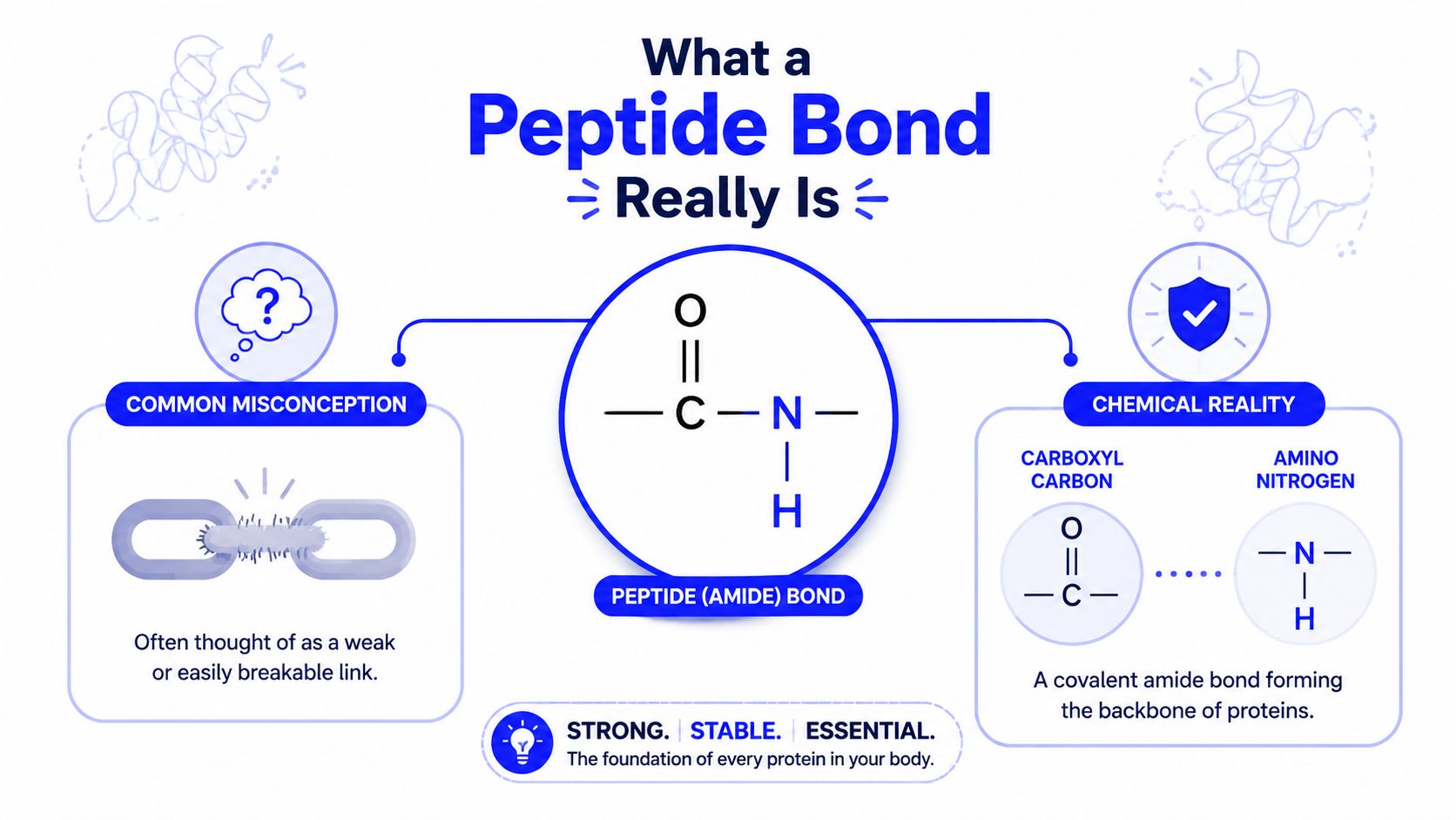
Students often picture a peptide bond as a vague “link” between amino acids. Chemically, it's much more specific: a peptide bond is a covalent amide bond formed between the carboxyl carbon of one amino acid and the amino nitrogen of another. That single definition identifies both atoms that matter and the connection that builds peptide chains.
A simplified backbone segment looks like this:
... Cα–C(=O)–N–Cα ...
The Cα, or alpha-carbon, is the central carbon of each amino-acid residue. It carries the side chain that gives alanine, serine, glycine, or another amino acid its chemical identity. The repeating C(=O)–N connection is the peptide bond, also called an amide linkage.
The vocabulary that prevents confusion
A peptide has direction. One end carries the free amino group and is called the amino terminus, or N-terminus. The other carries the free carboxyl group and is called the carboxyl terminus, or C-terminus. A chain is conventionally written from N-terminus to C-terminus, so the order of residues isn't decorative. Ala-Ser differs from Ser-Ala because the bond connects different functional groups in a different sequence.
Once an amino acid joins a chain, it's usually called a residue. The term reflects the fact that part of the original free amino acid has participated in bond formation. The side chains remain attached to their alpha-carbons and project from the repeating backbone.
Core idea: Every ordinary peptide bond uses the same amide chemistry. Protein diversity comes largely from the order and chemical properties of the side chains attached to the shared backbone.
Emil Fischer's synthesis of glycylglycine in 1901 provided one of the early experimental demonstrations that amino acids could be connected through amide bonds to form peptides, as described in this history of peptide chemistry. A dipeptide contains one such connection. A tripeptide contains two. Longer chains carry the same repeating pattern until folding brings the backbone into a three-dimensional structure.
How Peptide Bonds Form and Break
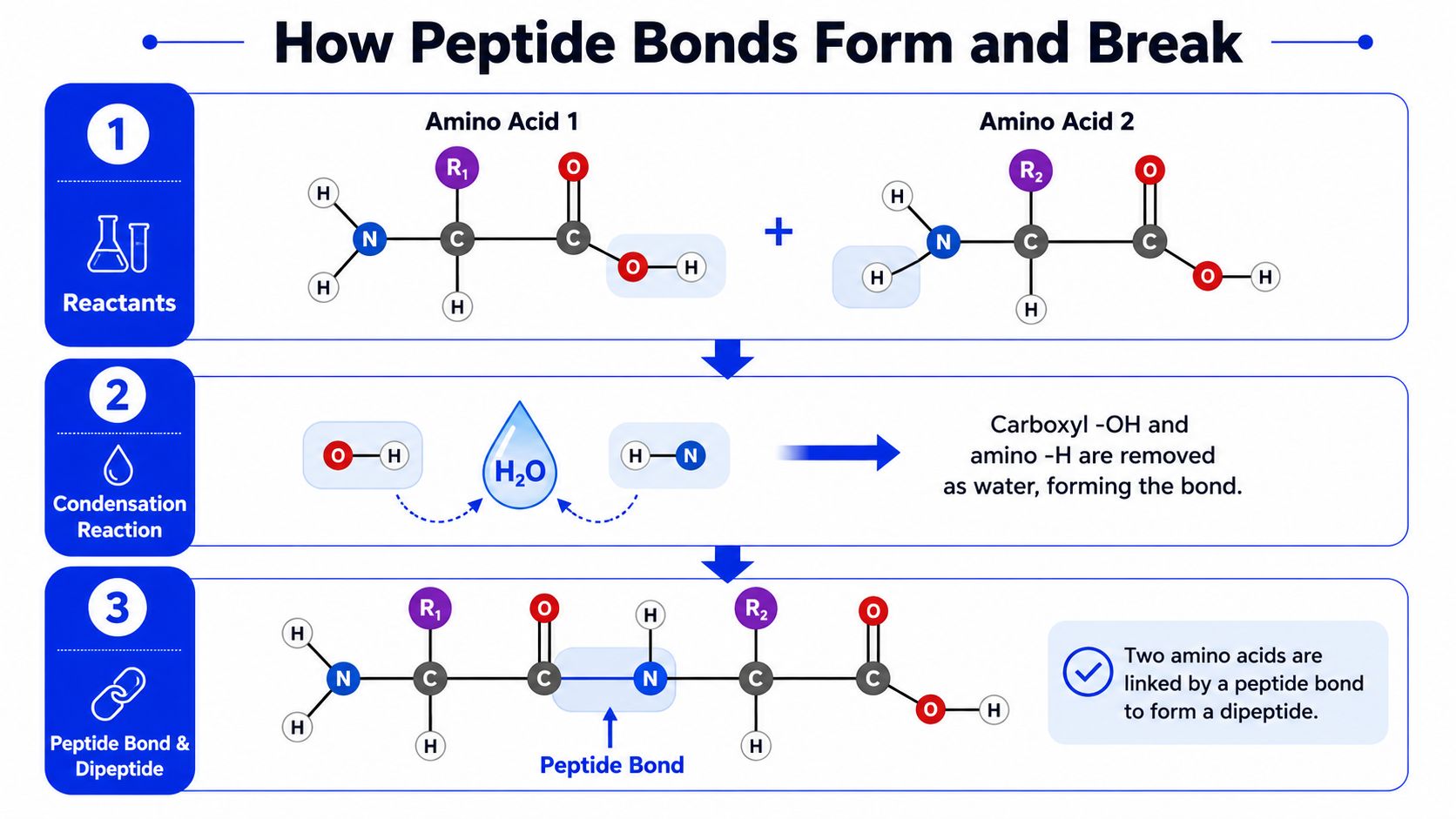
Peptide-bond formation is easiest to understand as a condensation reaction between two amino acids. Consider a generic first amino acid with a carboxyl group and a second amino acid with an amino group:
Amino acid 1–C(=O)–OH + H–N–Amino acid 2
The carboxyl group contributes its –OH. The amino group contributes a hydrogen. Those pieces combine as water, while the carboxyl carbon forms a new bond to the amino nitrogen:
Amino acid 1–C(=O)–OH + H–N–Amino acid 2 → Amino acid 1–C(=O)–N–Amino acid 2 + H₂O
The product is a dipeptide with an internal C(=O)–N amide linkage. The original amino group of the first residue remains the N-terminus, and the original carboxyl group of the second residue remains the C-terminus.
Formation requires chemical work
In a watery cellular environment, mixing amino acids doesn't efficiently build a long peptide. Cells use the ribosome and energy-dependent activation steps to direct bond formation during translation. Laboratory peptide synthesis also activates the reacting groups with coupling chemistry so the desired amide bond forms instead of remaining an unfavorable reaction in water.
The same connection can undergo the reverse reaction, hydrolysis. Water is consumed to split the amide linkage:
Amino acid 1–C(=O)–N–Amino acid 2 + H₂O → Amino acid 1–C(=O)–OH + H–N–Amino acid 2
Enzymes make this process selective. Proteases can cleave peptide bonds within proteins, while peptidases can process shorter peptides or trim residues from chain ends. Acidic or basic hydrolysis, often combined with heat, can break peptide chains more broadly for analytical work.
Why stability still has limits
A peptide bond is unusually stable under ordinary nonenzymatic conditions. One source reports a neutral-pH, 25°C half-life of about 350 to 600 years without enzymes, while structural measurements place the bond near 1.32 Å and show that cis peptide bonds occur in only about 0.03% of natively folded protein crystal structures, according to this peptide-bond data overview.
That stability doesn't mean a peptide is indestructible. Harsh pH, high temperature, enzymes, and contamination can promote cleavage or broader degradation. In a research workflow, protecting a peptide therefore means controlling both the molecule's chemistry and the conditions surrounding it.
Simple Examples of Peptide Bonds in Dipeptides and Tripeptides
Alanylserine, written Ala-Ser, is a useful first example because it keeps the structure small enough to count. Alanine's carboxyl group reacts with serine's amino group, producing one C(=O)–N linkage:
H₂N–CH(CH₃)–C(=O)–NH–CH(CH₂OH)–C(=O)–OH
The left side is the N-terminus, the right side is the C-terminus, and the central amide connection is the only peptide bond. The methyl side chain identifies alanine, while the hydroxymethyl side chain identifies serine. This is the key visual distinction: the backbone connection is chemically consistent, but side chains change the molecule's properties.
A tripeptide has three residues and two internal peptide bonds. Glutathione, commonly represented as γ-Glu-Cys-Gly, illustrates an important exception to oversimplified diagrams. Its glutamate-to-cysteine connection uses glutamate's side-chain carboxyl group, a gamma linkage, rather than the usual alpha-carboxyl arrangement. The molecule's peptide-bonded structure supports its role as an important cellular antioxidant.
Angiotensin II contains eight residues and seven internal peptide bonds. Its defined sequence produces a biologically active signaling molecule involved in vasoactive regulation. Bradykinin contains nine residues and eight internal peptide bonds, and its activity depends on preserving the correct sequence, because cleavage creates shorter fragments with different properties.
Oxytocin also contains nine residues and eight internal peptide bonds, but it's organized as a cyclic peptide through a disulfide bridge between cysteine residues. The disulfide bond is not a peptide bond. That distinction matters because the molecule contains two different kinds of covalent connections, one defining the amino-acid chain and another helping close its ring structure.
| Peptide | Residues | Peptide Bonds | Biological Role |
|---|---|---|---|
| Alanylserine | 2 | 1 | Structural example of a dipeptide |
| Glutathione | 3 | 2 | Cellular antioxidant |
| Angiotensin II | 8 | 7 | Vasoactive signaling peptide |
| Bradykinin | 9 | 8 | Signaling peptide whose activity depends on sequence integrity |
| Oxytocin | 9 | 8 | Cyclic signaling peptide with a disulfide bridge |
Counting the bonds becomes useful when preparing a peptide for research. For concentration calculations, sequence review, or solution preparation, researchers can use practical reconstitution help from Herbilabs alongside the molecule's documented sequence and mass.
Larger Examples of Peptide Bonds in Real Proteins
A protein is a long chain in which the peptide bond repeats through the backbone. The pattern can be written as:
Cα–C(=O)–N–Cα–C(=O)–N
Insulin makes the idea concrete because it contains two separate polypeptide chains. Its A chain has 21 residues and 20 peptide bonds, while its B chain has 30 residues and 29 peptide bonds. Disulfide bridges connect the chains, but those bridges aren't peptide bonds. The peptide bonds remain the links within each chain.
Lysozyme provides a contrasting example. It is a single chain with 129 residues and 128 peptide bonds. The difference between insulin and lysozyme shows why residue count alone doesn't reveal a protein's final shape or biological role. Chain arrangement, side-chain chemistry, disulfide connections, and noncovalent interactions all contribute to the folded structure.
| Protein | Residues | Peptide Bonds | Chains | Function |
|---|---|---|---|---|
| Insulin A chain | 21 | 20 | Part of a two-chain protein | Hormonal signaling |
| Insulin B chain | 30 | 29 | Part of a two-chain protein | Hormonal signaling |
| Lysozyme | 129 | 128 | Single chain | Enzymatic defense activity |
Sequence changes the folding landscape
Each peptide bond creates part of the backbone, but each residue also places a particular side chain beside that backbone. Hydrophobic groups may pack away from water. Charged groups may form ionic interactions. Polar groups can participate in hydrogen bonding. Cysteine residues can create disulfide bridges under suitable conditions.
Myoglobin and hemoglobin demonstrate the same principle in familiar oxygen-related proteins. Their biological functions differ because their sequences and assembled structures differ, not because one uses a fundamentally different type of peptide bond.
A protein's peptide bonds build the continuous scaffold. Its sequence determines how that scaffold can fold, move, bind, and function.
For a researcher reading a sequence, the first calculation is mechanical. A linear chain with a known residue count has one fewer internal peptide bond than residues. The deeper question is structural: which side chains surround those bonds, and what three-dimensional arrangement does that sequence favor?
Why Peptide Bond Structure Shapes Protein Behavior
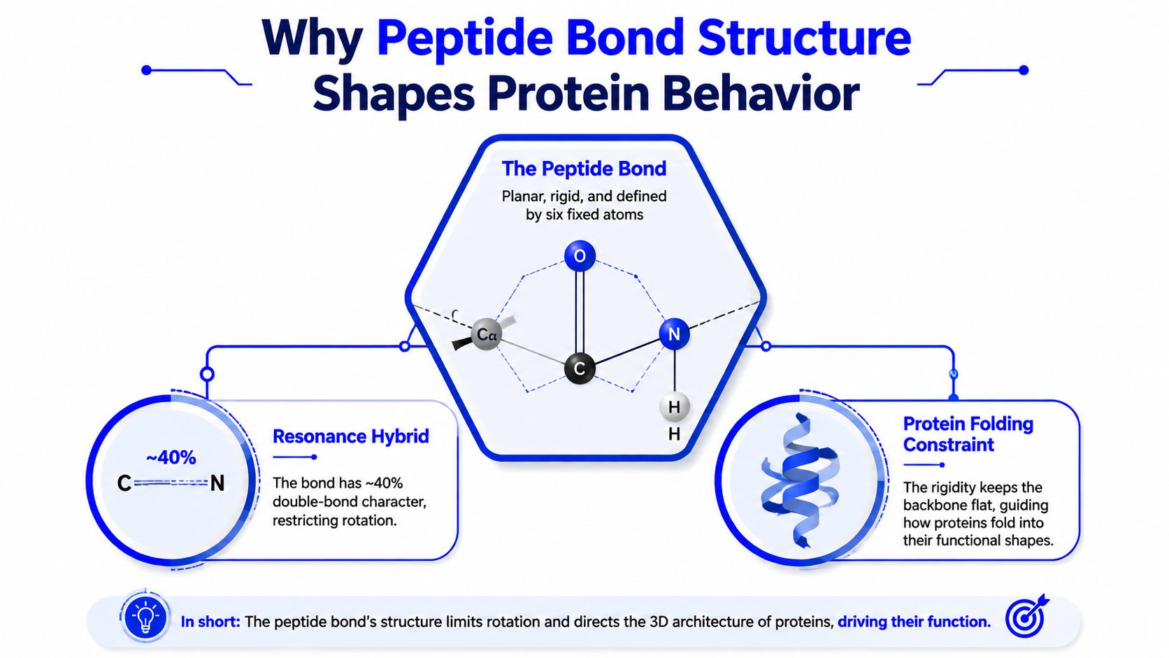
The peptide C–N bond isn't a freely rotating single bond. Resonance allows electron density to spread between the carbonyl group and the nitrogen, giving the linkage partial double-bond character. Structural measurements place the peptide C–N bond at about 1.32 Å, between a typical C–N single bond at 1.49 Å and a C=N double bond at 1.27 Å, as summarized in this biochemistry treatment of peptide-bond structure.
A rigid unit inside a flexible chain
The partial double-bond character restricts rotation around the C–N connection. The carbonyl carbon, carbonyl oxygen, amide nitrogen, and the neighboring alpha-carbons occupy a largely planar arrangement. The chain still has flexibility because rotation can occur around adjacent bonds, but the peptide bond itself acts more like a stiff panel than a loose hinge.
Most peptide bonds favor the trans arrangement, which places neighboring alpha-carbons on opposite sides of the amide linkage. Cis arrangements are possible, but they're uncommon in folded proteins. This balance between rigid peptide units and rotation at nearby bonds gives a chain enough structure to fold without behaving like a completely fixed rod.
Folding and molecular design
Alpha-helices and beta-sheets depend on repeated backbone geometry and hydrogen bonding. The peptide bond supplies the carbonyl oxygen and amide hydrogen involved in those interactions, while the restricted geometry limits which conformations the chain can adopt.
Local structure can also influence the electronic character of peptide bonds. A 2025 review and commentary in IUCrJ discusses evidence that peptide bonds in alpha-helical environments can appear more enol-like than those in beta-strands and emphasizes the need for high-resolution experimental structures to clarify these differences. That nuance addresses a question basic explanations often miss: peptide bonds share core amide chemistry, but their local structural environments can affect how that chemistry is expressed. The discussion appears in this open-access structural review.
Drug designers use this structural knowledge when developing peptidomimetics, amide isosteres, and beta-amino-acid-based compounds. These designs can alter backbone geometry or reduce susceptibility to enzymatic cleavage while preserving useful recognition features. The aim isn't to discard peptide-bond chemistry. It's to adjust the parts of the molecular framework that control stability, shape, and biological handling.
Peptide Bonds in the Research Workflow
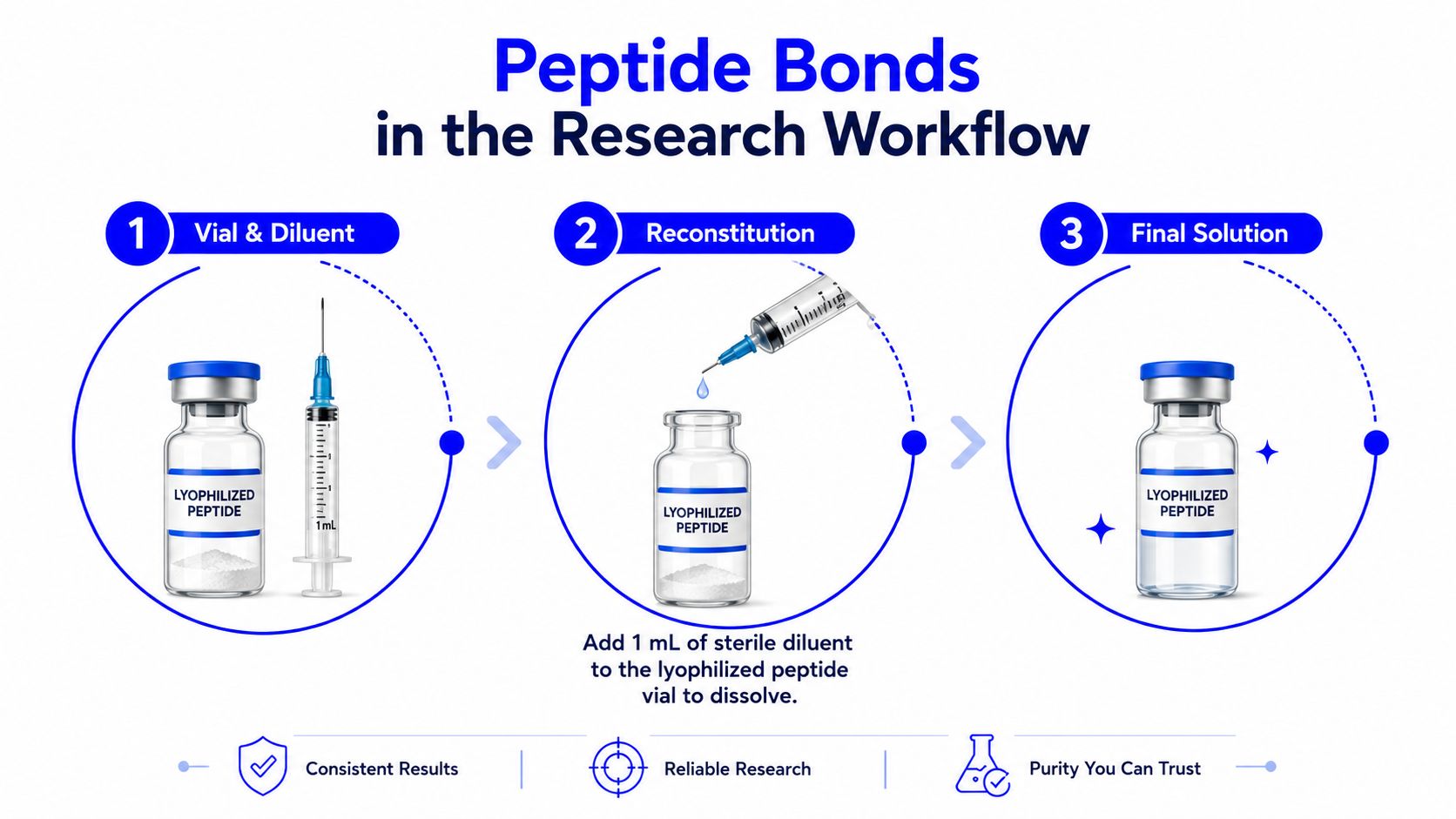
A lyophilized peptide vial turns the same chemistry into a handling problem. The peptide bonds are already present, but the research team must restore the material to solution without introducing contamination, creating unnecessary stress, or losing track of concentration.
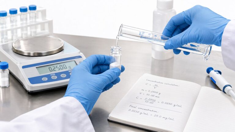
A typical workflow begins with the product documentation. The researcher checks the peptide mass, intended diluent, solubility guidance, and required final concentration before selecting a sterile diluent and volume. The arithmetic is simple, but the record must distinguish total peptide mass from final solution volume.
Aseptic handling protects the sample
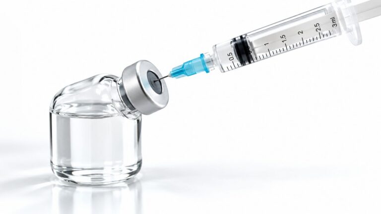
Bacterial contamination matters because microorganisms can introduce proteolytic enzymes that cleave peptide bonds. A controlled bench routine therefore includes the following actions:
- Prepare the workspace: Organize the vial, sterile diluent, syringe, needle, labels, and waste container before opening anything.
- Clean the stopper: Wipe the vial septum with 70% isopropanol and allow it to dry.
- Use fresh sterile components: A new sterile needle and syringe should be used for each vial to reduce cross-contamination.
- Add liquid gradually: Direct the diluent against the vial wall when possible, rather than forcefully striking the powder.
- Mix gently: Swirling can dissolve the material while avoiding the unnecessary agitation associated with vigorous vortexing.
- Label and aliquot: Record concentration and preparation details, then divide the solution when repeated access would create avoidable freeze-thaw exposure.
For researchers comparing suppliers, Herbilabs offers a sterile Reconstitution Solution in multi-dose glass vials for research use, described for preparing working solutions from lyophilized peptides, proteins, and antibodies. The product information includes sterile, non-pyrogenic solution specifications, lot-specific documentation, and an in-use period of up to 28 days when proper aseptic technique is used. A separate step-by-step peptide reconstitution guide can support the operational planning, while the peptide's own documentation remains the controlling source for compatibility and storage.
Common Questions About Peptide Bonds in the Lab
1. How can a researcher count peptide bonds?
For a linear chain, subtract one from the number of residues. Two residues give one internal peptide bond. Three residues give two. A chain with a known sequence can also be inspected directly by locating each –C(=O)–N– connection between neighboring residues.
The shortcut needs adjustment for unusual structures. Cyclic peptides don't have free ends, and side-chain linkages, such as the gamma connection in glutathione, require attention to the actual structure rather than relying only on a standard alpha-backbone diagram. Disulfide bridges must be counted separately because they aren't peptide bonds.
2. What does hydrolysis do?
Hydrolysis uses water to cleave an amide linkage. Depending on the conditions, a chain can break into two shorter fragments or continue toward free amino acids. Enzymatic hydrolysis uses proteases or peptidases with substrate preferences, while strongly acidic or basic conditions can promote broader chemical cleavage.
That distinction matters during analysis. A researcher investigating sequence integrity must separate intentional digestion from accidental degradation caused by unsuitable pH, heat, enzymes, or contamination.
3. Why do storage and reconstitution affect integrity?
Lyophilized material is generally easier to preserve than a prepared solution, but the supplier's instructions remain decisive. Once a peptide is in solution, researchers should use a documented diluent, minimize repeated vial access, aliquot when appropriate, and avoid unnecessary freeze-thaw cycles.
A bacteriostatic diluent may support multi-dose research handling when used aseptically, but it doesn't eliminate the need for labeling, controlled storage, and compatibility checks. Guidance on bacteriostatic water can help organize handling decisions, while the specific peptide documentation should determine the final workflow.
| Question | Quick Answer |
|---|---|
| How many peptide bonds are in a linear chain? | One fewer than the number of residues |
| What is hydrolysis? | Water-mediated cleavage of an amide linkage |
| Are disulfide bridges peptide bonds? | No, they're a separate type of covalent connection |
| What protects a prepared peptide solution? | Aseptic handling, suitable storage, aliquoting, and limited freeze-thaw exposure |
Understanding peptide bonds becomes much easier when every example returns to the same structural test: identify the carbonyl carbon, identify the amino nitrogen, and locate the C(=O)–N amide linkage. That method works for a dipeptide, a hormone, or a protein backbone.
For research teams preparing lyophilized peptides, proteins, or antibodies, Herbilabs provides sterile reconstitution solutions, multi-dose vial formats, and lot-specific quality documentation for RUO workflows. Visit Herbilabs to review available diluents and reconstitution resources before setting up the next laboratory preparation.